Filamentous microbes, cellular and molecular aspects of host-microbe interactions.
Born in Cannes (France) and trained in Nice (France), Dr. Edouard Evangelisti carried out postdoctoral research at the Sainsbury Laboratory, University of Cambridge (UK), before serving as Assistant Professor and group leader at Wageningen University (the Netherlands). Since May 2024, he has held a Chair of Excellence (IDEX UCA-JEDI) at Université Côte d’Azur.
Understanding how filamentous microorganisms colonize plants
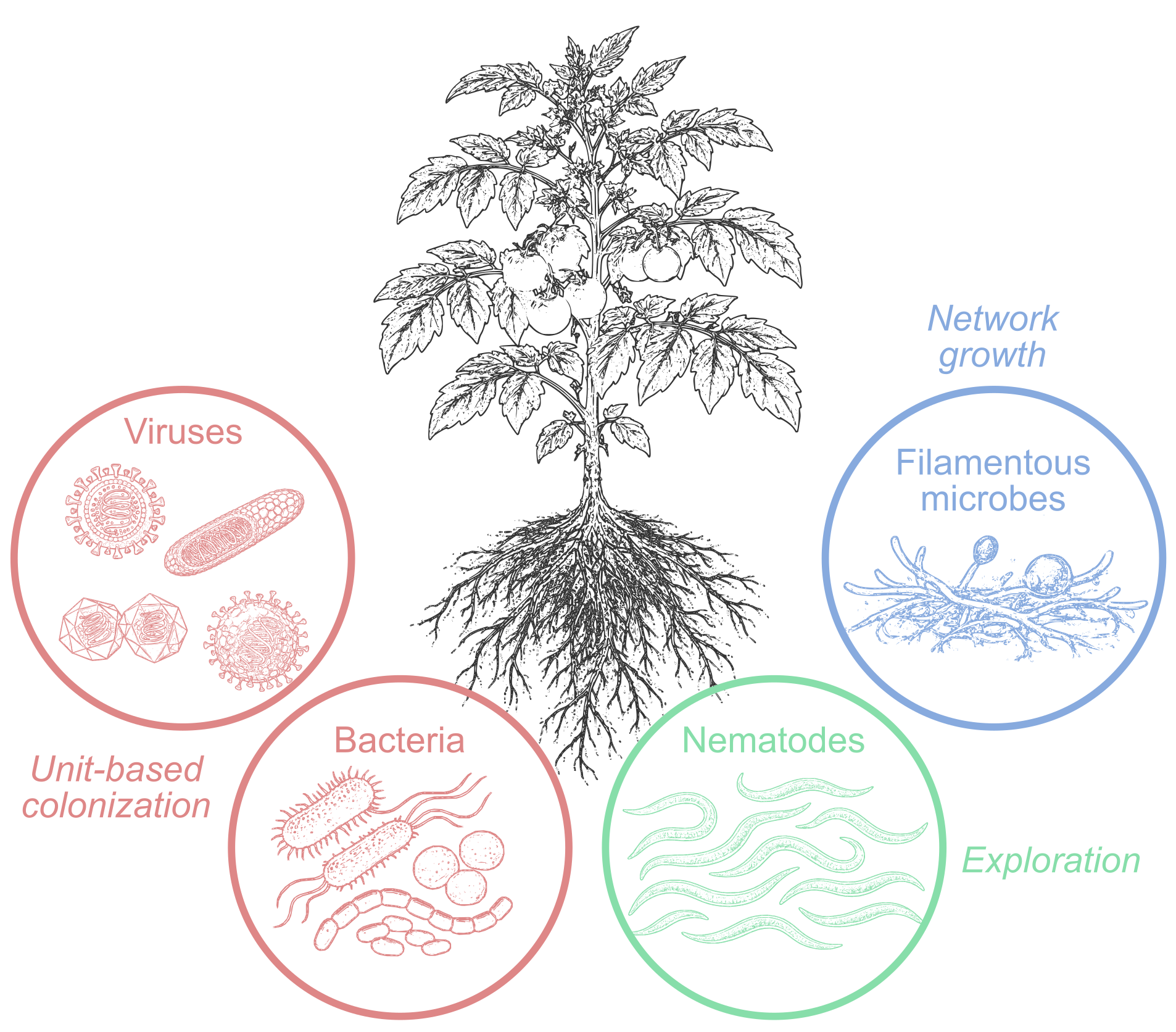
My research aims to decipher how filamentous microorganisms, in particular oomycetes, colonize plant tissues and interact with their host. In contrast to viruses and bacteria, which colonize through the proliferation of discrete units, and to nematodes, which explore tissues through active movement, oomycetes develop as continuous networks. Starting from a single spore, they can extend their mycelium over distances several orders of magnitude greater than their initial size, while deploying specialized structures for host interaction and successive genetic programs. This distinctive spatial organization raises a central question: how do these systems coordinate their activities across multiple scales, from the cellular level to the plant tissue? My approach combines molecular biology, cell biology, imaging, and computational analysis to identify the fundamental principles governing this colonization.
Modes of plant colonization by microorganisms. From left to right: strategies based on the proliferation of discrete units (viruses, bacteria), active exploration (nematodes), or the extension of continuous networks (filamentous microorganisms).
Perception and subcellular organization in filamentous microorganisms
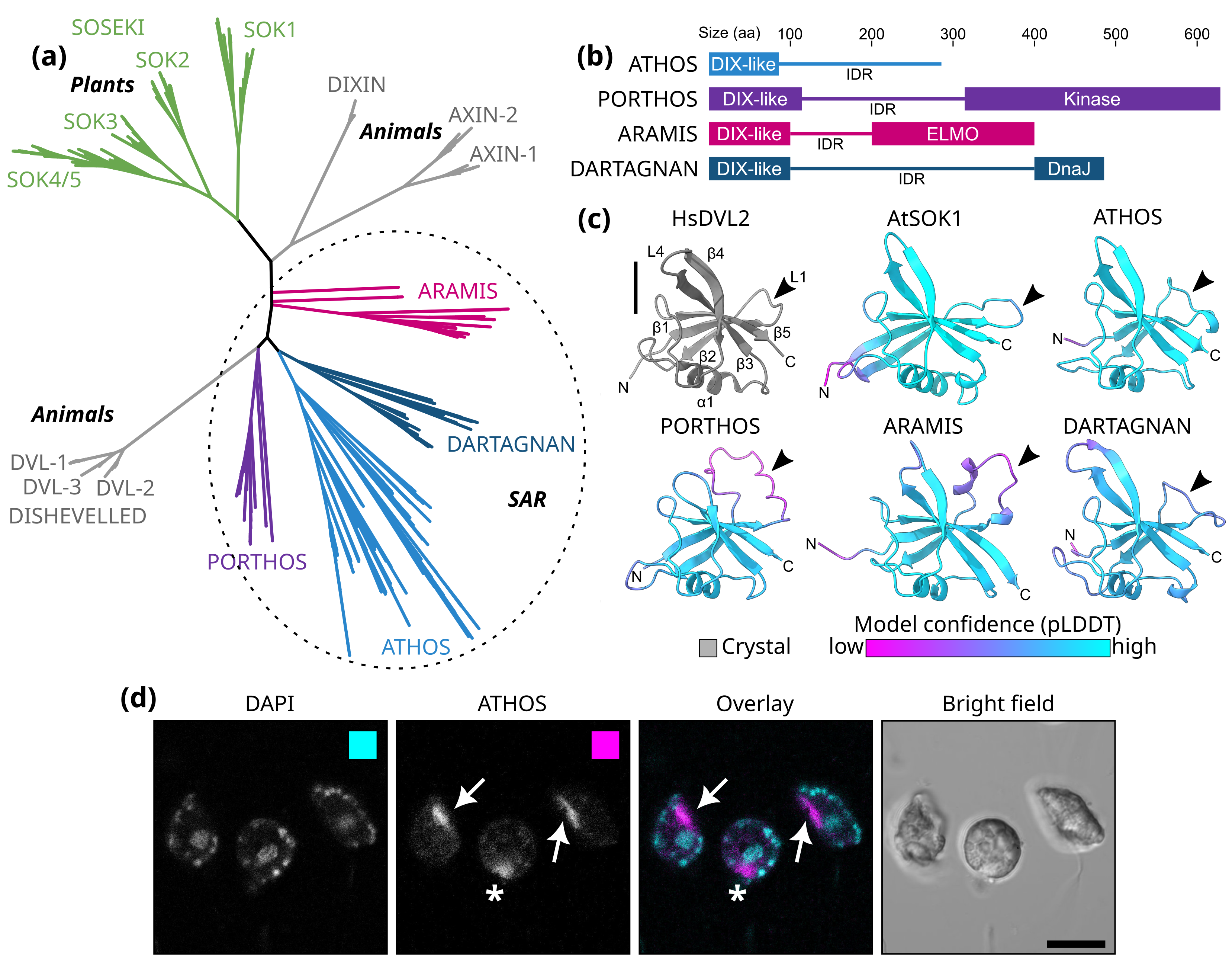
This research axis aims to establish a conceptual framework to understand how filamentous microorganisms perceive their environment and organize their growth at the subcellular scale. It is based on the idea that the colonization of plant tissues does not rely solely on genetic programs, but also on the ability of cells to integrate physical constraints and to organize into functional compartments, independent of classical organelles. This organization indicates that the perception and integration of such constraints play a central role in shaping the functional architecture of filamentous cells. My work has led to the identification of a new family of DIX domain-containing proteins, specific to certain eukaryotes within the SAR supergroup. In Phytophthora, these proteins display a highly specific localization within a subcellular compartment of zoospores, suggesting intrinsic self-organizing properties, likely linked to the combination of a DIX domain and intrinsically disordered regions. These findings support a model in which dynamic protein assemblies contribute to the internal structuring of cells and to the integration of environmental signals. They reveal principles of cellular organization that are distinct from established paradigms in animals and plants, and position filamentous microorganisms as model systems for studying cellular self-organization.
Identification and characterization of a family of DIX domain-containing proteins in eukaryotes of the SAR supergroup (a), likely forming molecular assemblies (b-c) and localized to a specific subcellular compartment in Phytophthora zoospores (d). Adapted from Kostareli et al. (2025).
Molecular interactions and dynamics of plant tissue colonization
A second research axis aims to understand how filamentous microorganisms interact with their host and structure tissue colonization at the molecular and cellular levels. My work has shown that these organisms secrete proteins capable of hijacking plant cellular processes, targeting key functions such as intracellular trafficking and signaling networks. These strategies are not restricted to pathogens: similar mechanisms are also employed by symbiotic microorganisms, suggesting the existence of a functional continuum between beneficial and pathogenic interactions.
Building on these findings, I approach colonization as a spatiotemporally organized process, involving the dynamic distribution of effectors, the reconfiguration of plant cellular structures, and the emergence of specialized interaction interfaces. This perspective complements my first research axis by linking the organizational principles of microorganisms to their ability to remodel their cellular environment. To capture this complexity, I develop quantitative approaches based on artificial intelligence, enabling the transformation of microscopic observations into measurable data. Tools such as AMFinder (https://github.com/SchornacklabSLCU/amfinder) and HFinder (https://github.com/EEvangelisti/hfinder) allow automated analysis of plant tissue colonization and the quantification of key parameters, including effector localization, organelle reorganization, and the accumulation of immune receptors at infection interfaces. By moving from qualitative observation to systematic and reproducible description, these approaches establish a quantitative framework for studying colonization. The ultimate goal is to develop predictive models capable of anticipating microbial behavior in complex environments.
Use of deep learning to extract and quantify fungal structures (hyphae) and plant-pathogen interfaces (haustoria), as well as plant organelles (nuclei, chloroplasts). These spatially resolved data transform microscopic observations into a quantitative description of colonization, paving the way for predictive models of growth within plant tissues. Adapted from Korovesis et al. (2026).
An ambition: building predictive models of colonization
My research aims to establish an integrated understanding of how filamentous microorganisms colonize plant tissues, by linking molecular mechanisms, cellular organization, and the spatial dynamics of mycelial growth. The goal is to develop predictive models capable of anticipating the growth, differentiation, and infection behavior of these organisms in complex environments. These models will integrate key factors such as interactions with the microbiome, microbial competition, and tissue-level constraints, in order to describe and predict infection dynamics in situ. Ultimately, this approach aims to better forecast pathogen behavior in soils and to inform breeding strategies to address soil-borne diseases. By linking fundamental mechanisms with predictive modeling of infection dynamics, this research contributes to the agroecological ambitions of the Sophia Agrobiotech Institute and INRAE, particularly in the management of soil-borne diseases, where current approaches remain limited.
International collaborations
Affiliated with the GreenTE consortium (https://green-te.nl/), which investigates how plant cells perceive mechanical forces and how these regulate growth, development, and immunity.
Member of the steering committee of the Oomycete Molecular Genetics Network (OMGN), where I oversee the development and maintenance of the website (https://oomycetes.com/).
Co-supervisor, with Dr. Nicolas Desneux (MIB team), of two PhD students from the Beijing Academy of Agriculture and Forestry Sciences (BAAFS), within the framework of an international partnership between BAAFS and Université Côte d’Azur.
Chair of Excellence IDEX UCA-JEDI 2024-2027. Relentless pathogens: how do they sustain growth, attack hosts and outcompete other microbes. Funded by LABEX SIGNALIFE ANR-11-LABX-0028-01 and IDEX UCAJedi ANR-15-IDEX-01.
Recent publications
Korovesis S, Wang S, Xu L, Giraudon I, Rosales Hernandez D, Panek E, Boeglin L, Kostareli MM, Pluis MHJ, Wang B, Wang Y, Abdennour D, Keller H, Birch PRJ, Schornack S, Evangelisti E2026. Deep learning enables quantitative subcellular analysis of plant-microbe interfaces. BioRxiv.
Kostareli MM, Westerink T, Couillaud G, Peippo M, Govers F, Weijers D, Evangelisti E. 2025. Diversification of DIX domain-containing proteins in the SAR supergroup. mBio: e0396624. Yuen ELH, Tumtas Y, King F, Ibrahim T, Chan LI, Evangelisti E, Tulin F, Skłenar J, Menke FLH, Kamoun S, et al.2024. A pathogen effector co-opts a host RabGAP protein to remodel pathogen interface and subvert defense-related secretion. Science advances10: eado9516. Teulet A, Quan C, Evangelisti E, Wanke A, Yang W, Schornack S. 2023. A pathogen effector FOLD diversified in symbiotic fungi. New Phytologist239: 1127–1139.
Evangelisti E, Guyon A, Shenhav L, Schornack S. 2023. FIRE mimics a 14-3-3-binding motif to promote Phytophthora palmivora infection. Molecular Plant-Microbe Interactions36: 315–322. Evangelisti E, Turner C, McDowell A, Shenhav L, Yunusov T, Gavrin A, Servante EK, Quan C, Schornack S. 2021. Deep learning-based quantification of arbuscular mycorrhizal fungi in plant roots. The New Phytologist232: 2207–2219.
By browsing our site you accept the installation and use cookies on your computer.
Know more
About cookies
What is a "cookie"?
A "cookie" is a piece of information, usually small and identified by a name, which may be sent to your browser by a website you are visiting. Your web browser will store it for a period of time, and send it back to the web server each time you log on again.
Different types of cookies are placed on the sites:
Cookies strictly necessary for the proper functioning of the site
Cookies deposited by third party sites to improve the interactivity of the site, to collect statistics
Cookies strictly necessary for the site to function
These cookies allow the main services of the site to function optimally. You can technically block them using your browser settings but your experience on the site may be degraded.
Furthermore, you have the possibility of opposing the use of audience measurement tracers strictly necessary for the functioning and current administration of the website in the cookie management window accessible via the link located in the footer of the site.
Technical cookies
Name of the cookie
Purpose
Shelf life
CAS and PHP session cookies
Login credentials, session security
Session
Tarteaucitron
Saving your cookie consent choices
12 months
Audience measurement cookies (AT Internet)
Name of the cookie
Purpose
Shelf life
atid
Trace the visitor's route in order to establish visit statistics.
13 months
atuserid
Store the anonymous ID of the visitor who starts the first time he visits the site
13 months
atidvisitor
Identify the numbers (unique identifiers of a site) seen by the visitor and store the visitor's identifiers.
13 months
About the AT Internet audience measurement tool :
AT Internet's audience measurement tool Analytics is deployed on this site in order to obtain information on visitors' navigation and to improve its use.
The French data protection authority (CNIL) has granted an exemption to AT Internet's Web Analytics cookie. This tool is thus exempt from the collection of the Internet user's consent with regard to the deposit of analytics cookies. However, you can refuse the deposit of these cookies via the cookie management panel.
Good to know:
The data collected are not cross-checked with other processing operations
The deposited cookie is only used to produce anonymous statistics
The cookie does not allow the user's navigation on other sites to be tracked.
Third party cookies to improve the interactivity of the site
This site relies on certain services provided by third parties which allow :
to offer interactive content;
improve usability and facilitate the sharing of content on social networks;
view videos and animated presentations directly on our website;
protect form entries from robots;
monitor the performance of the site.
These third parties will collect and use your browsing data for their own purposes.
How to accept or reject cookies
When you start browsing an eZpublish site, the appearance of the "cookies" banner allows you to accept or refuse all the cookies we use. This banner will be displayed as long as you have not made a choice, even if you are browsing on another page of the site.
You can change your choices at any time by clicking on the "Cookie Management" link.